 ISSN : 1598-2939
© Korea Institute of Sport Science
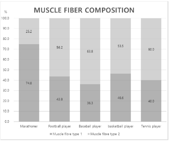
The objective of the present study was to assess muscle fibre composition by non-invasive methods for various sports events. 53 males of marathon (n=5), football (n=22), baseball (n=14), basketball (n=8) and tennis (n=4) athletes volunteered to participate in this study. The following 7 tests (overhead medicine ball throw, pull up, standing broad jump, sit up, shuttle run 10 x5, push up, vertical jump) are relatively easy to perform and to gather information about fibre composition through fitness levels. It can be used to assess the performance of strength using standard tables and to receive an indication of the degree of their share of fast (type II) and slow (type I) muscle fibres. For the study, a data analysis program in Microsoft Excel was used to view the mean and standard deviation of muscle fibre types. As a result, it was possible to speculate that the ratio of muscle fibre types differed according to the type of exercise. The results were obtained after sport motor tests of the mean value of muscle fibre composition by different sports events. Fibre type distribution remained with about 74.8% type 1 and 25.2% type 2 by marathoners, 43.8% type 1 and 53.2% type 2 by football athletes, 36.3% type 1 and 63.8% type 2 by baseball athletes, 46.6% type 1 and 53.5% type 2 by basketball athletes, and 40.0% type 1 and 60.0% type 2 by tennis athletes. Through these tests, the direction of the athletes’ muscle development can be considered, and it is possible to check the muscle fibre composition of elite athletes for the improvement of performance for each sport event. Moreover, it assumes that if this test is used for children, adolescents and young athletes, it will help to design scientific and effective training programs, thereby improving athletes’ ability to perform.
The objective of the present study was to assess muscle fibre composition by non-invasive methods for various sports events. 53 males of marathon (n=5), football (n=22), baseball (n=14), basketball (n=8) and tennis (n=4) athletes volunteered to participate in this study. The following 7 tests (overhead medicine ball throw, pull up, standing broad jump, sit up, shuttle run 10 x5, push up, vertical jump) are relatively easy to perform and to gather information about fibre composition through fitness levels. It can be used to assess the performance of strength using standard tables and to receive an indication of the degree of their share of fast (type II) and slow (type I) muscle fibres. For the study, a data analysis program in Microsoft Excel was used to view the mean and standard deviation of muscle fibre types. As a result, it was possible to speculate that the ratio of muscle fibre types differed according to the type of exercise. The results were obtained after sport motor tests of the mean value of muscle fibre composition by different sports events. Fibre type distribution remained with about 74.8% type 1 and 25.2% type 2 by marathoners, 43.8% type 1 and 53.2% type 2 by football athletes, 36.3% type 1 and 63.8% type 2 by baseball athletes, 46.6% type 1 and 53.5% type 2 by basketball athletes, and 40.0% type 1 and 60.0% type 2 by tennis athletes. Through these tests, the direction of the athletes’ muscle development can be considered, and it is possible to check the muscle fibre composition of elite athletes for the improvement of performance for each sport event. Moreover, it assumes that if this test is used for children, adolescents and young athletes, it will help to design scientific and effective training programs, thereby improving athletes’ ability to perform.
The human skeletal muscle consists of different types of fibre. Studies by Campbell (1979) (Campbell, Bonen, Kirby, & Belcastro, 1979) revealed the first physiological characteristics of the fibre types. This researcher found both color (red, slow and white, fast muscle fibre) and contractile properties.
In human skeletal muscles, two types of fibre can be distinguished based on light microscopic histochemical, biochemical, electrophysiological considerations (Gollnick & Matoba, 1984). Slow muscle fibres (ST or type 1 fibre) contract and relax relatively slowly (contraction times approx. 80ms). These fibres have a high myoglobin content, which enables rapid intracellular O2 transport (Gollnick & Matoba, 1984). Fast muscle fibres (FT or type 2 fibre) take a relatively short time to develop maximum tension (approx. 30ms). Their ability to contract quickly is based, among other things, on their high myosin ATPase activity (Gollnick & Matoba, 1984).
Human skeletal muscles consist of different percentages of these types of fibre. This percentage increase varies widely between different muscle types and between different individuals (Gollnick et al., 1974). It appears that exercise can induce muscle fibre transformation and alter muscle fibre composition (Staron & Johnson, 1993).
Regarding endurance training, studies have shown that there is an increase in type 1 fibre and a decrease in type 2b fibre (Henriksson & Hickner, 1994; Howald et al., 1985). Other studies showed that after strength training there was a shift in muscle fibre within the type 2 fibre from type 2b to type 2a fibre in both men and women (Wang et al., 1993). e more athletes know about their muscle fibre types, the better they can train and a successful school athlete could describe a genetic distribution of the fibre types regarding a suitable sport. Moreover, school athletes can improve performance early according to individual ability (Colliander et al., 1988). However, medical and human medical studies require approval from an ethics committee. In Korea, of course, this also applies to those research projects in which muscle biopsies are performed. The question arises to what extent the removal of muscle tissue from the living is ethical (Leitner, 2009). The discussion mainly refers to the biopsy of healthy volunteers and less to the tissue extraction diagnosis of any muscular disorders (Edwards et al., 1983). If there is a disease, the patient must be subjected to such an intervention, as this can cause severe, sometimes deadly diseases (polymyositis), which can be diagnosed (Edwards et al., 1983). However, when working with healthy, volunteer subjects for research purposes, it is not so easy to justify (Dietrichson et al., 1987). It is therefore of importance here that a tissue extraction technique is used that places the smallest possible strain on the test subject and leaves no permanent damage in the long term.
For this reason, since 1995, the Federal Institute of Sport Science in Germany applies a test to determine muscle fibre composition with many sport motoric tests in school (Beck, 1995). The non-invasive muscle fibre composition tests are one of many easy-to-use instruments. Nevertheless, to the authors’ knowledge, no study has reported the calculation of muscle fibre distribution with non-invasive methods among different sport events. Therefore, the objective of the present study was to find the calculation of muscle fibre composition with non-invasive tests for various sports events.
53 males of marathon (n=5), football (n=22), baseball (n=14), basketball (n=8) and tennis (n=4) K University athletes in Seoul volunteered to participate in this study. Subjects were athletes (mean ± SD, age: 20.4±1.3) and they have no chronic and acute pain. All subjects had previously been screened and diagnosed by an orthopaedic surgeon. Subjects did not present any neurological signs of pathological importance in the clinical examination. Prior to the study, participants were informed about the purpose, procedures and risks of the study and written informed consent was obtained from each participant.
Figure 1 shows the systematization of motor skills according to Bös (Bös et al., 2009). This distinction serves as a basis for the development and classification of diagnostic procedures fort the assessment of motor performance. On a first level, motor skills are differentiated into energetically determined (conditional) skills and information-oriented (coordinative) skills. The relevant complex tests for recording these ability complexes are called coordination tests. On a second level, a distinction is made between the central ability categories endurance, strength, speed, coordination and mobility. On a more detailed third level, 10 ability components (AE: aerobic endurance, AnE: anaerobic endurance, SE: strength endurance, MS: maximum strength, HS: high-speed strength, AS: action speed, RS: reaction speed, CTP: coordination under time pressure, CPT: coordination for precision tasks, M: mobility) can be distinguished on the basis of stress norms (Bös et al., 2009).
Selected 7 tests (Tab. 1) are popular and simply test methods to measure motor skills in the school in Germany. The tests are easy to assessment fitness level and fiber composition for each student. The number of tests 1, 3, 5 and 7 are for explosive strength as Type 2 fiber and number of tests 2, 4, 6 are for strength endurance as Type 1.
The following test is easy to perform and to obtain information about fibre composition through fitness levels. You can use it to assess the performance of your strength using standard tables and receive an indication of how large their share of fast (type II) and slow (type I) muscle fibre is (Table 1).
| NO of test | Test | Point of assessment | ||||
|---|---|---|---|---|---|---|
| 4 | 3 | 2 | 1 | 0 | ||
| 1 | Overhead medicine ball throw test (m) | |||||
| 2 | Pull up (sec) | |||||
| 3 | Standing broad jump (cm) | |||||
| 4 | Sit up (30sec) | |||||
| 5 | Shuttle run 10 x5 (sec) | |||||
| 6 | Push up (30sec) | |||||
| 7 | Vertical jump (cm) | |||||
| Sum of test from 1, 3, 5, 7 for FT |
||||||
| Sum of test from 2, 4, 6 for ST |
||||||
In addition, Table 2 Table shows the formula is calculating the proportions of fast and slow twitch muscle fibre. It was calculated as follows X is the sum of test 1, 3, 5 and 7 and Y is the sum of test 2, 4 and 6. Percentage of fast twitch muscle fibers is calculated 100% - ( Y / (X + Y) * 100) and percentage of slow twitch muscle fibers is calculated 100% - ( X / (Y + X) * 100).
| Percentage of fast twitch muscle fibre = 100 - (Y / (X + Y) x 100) |
| Proportion of slowly twitching muscle fibre = 100 – (X / (X + Y) x 100) |
| X = sum of exercise 1, 3, 5 and 7 |
| Y = sum of exercise 2, 4 and 6 |
| Fast muscle fibre composition _____% Slow muscle fibre composition _____% |
Table 3 shows the standard values of sport motor tests according to Beck & Bös (Bös et al., 2009).
| Test | 4 Point | 3 Point | 2 Point | 1 Point | 0 Point |
|---|---|---|---|---|---|
| Excellent | Good | Normal | Under | Poor | |
| 7. Vertical jump (cm) | > 62 | 56 – 62 | 49 – 55 | 42 – 48 | < 42 |
From a parallel stand, with a double-armed throw-off, throw a 2kg medicine ball as far forward as possible. The test person stands with both feet in front of the throw-offline; the feet are hip-wide apart. When throwing the ball backwards, the body can be swung backwards, but the foot position must not be changed. During the test, the feet must not leave the ground, lifting the heels is allowed. They have 3 executions and the best result is counted.
The test person hangs on the bar shoulder-wide with both hands, thumbs should grasp the bar. With the support of the test leader or another person, the test person pulls the body up until the tip of the chin touches the bar. Immediately after the chin-up position is reached, the test leader removes himself and starts the stopwatch. He should stay in close proximity to the test person to be able to assist him if necessary and avoid injury.
The test person stands parallel to the front edge of the jump line. He decides independently about the time of the jump. Swinging with the arms and by bending the knees is allowed. The jump takes place on both legs. Landing is on both feet; it may not be grabbed backwards with the hand. They have 3 executions and the best result is counted.
The test person lies in a supine position on a gymnastics mat. The knees are bent (90 degrees). The hands are crossed at the neck. A partner kneels in front of the test person and fixes his feet (on the instep) to the floor. On the command "Ready-Lose" the test person straightens up and touches the knees with the elbows. Then they return to the starting position. This exercise is repeated as often as possible in the given time
The test person stands behind the start/finish line in the high start position. The run is started with the command "Finish-Lot". At the turns at the start/finish line and the turn mark, one foot must touch or cross the line. At the finish line, one foot must cross the finish line.
The hands are supported directly under the shoulders, fingers pointing forward. The upper body and legs remain stretched, the head in a straight extension of the spine. The arms are bent to a 90 ° angle and the body is lowered. Hips must not be bent. Then the subject pushes up to the starting position.
Measurements were completed using an electronic jumping mat (Kyunghee-Sport-Industrie, KHS-119). The test person stands barefoot on the mat with both legs, shoulder-width apart and body weight equally distributed between the both legs. The subjects squatted down to a ~90° knee flexion before beginning a powerful upward motion. The participants were ordered to jump as high as possible and the athletes were requested not to flex their knees to the highest level. They have executions and the best result is counted.
For the study data to analysis program in Microsoft Excel was used to view the mean and standard deviation of muscle fibre types and effect size and statistical power for this study in Overhead medicine ball throw test, Pull up, Standing broad jump, Sit up, Shuttle run 10 x5, Push up and Vertical jump were analysed F-test, ANOVA standard deviations of differences using G*Power power (α = 0.97) analysis program (Cohen, 1988; Faul et al., 2009).
Table 4 shows the roh-data of individual values and Figure 2 shows the result of mean values of ratio of muscle fibre type 1 and 2 composition.
| Sports event | subject | Sum of FT (score) |
Sum of ST (score) |
Sum of FT (%) |
Sum of ST (%) |
|---|---|---|---|---|---|
| Marathoner | R1 | 1 | 8 | 11.1 | 88.9 |
| R2 | 6 | 9 | 40 | 60 | |
| R3 | 2 | 10 | 16.7 | 83.3 | |
| R4 | 3 | 9 | 25 | 75 | |
| R5 | 5 | 10 | 33.3 | 66.7 | |
| Foot Ball | F1 | 10 | 10 | 50 | 50 |
| F2 | 12 | 10 | 54.5 | 45.5 | |
| F3 | 9 | 9 | 50 | 50 | |
| F4 | 12 | 10 | 54.5 | 45.5 | |
| F5 | 6 | 10 | 37.5 | 62.5 | |
| F6 | 12 | 11 | 52.2 | 47.8 | |
| F7 | 8 | 10 | 44.4 | 55.6 | |
| F8 | 11 | 10 | 52.4 | 47.6 | |
| F9 | 10 | 11 | 47.6 | 52.4 | |
| F10 | 14 | 10 | 58.3 | 41.7 | |
| F11 | 16 | 7 | 69.6 | 30.4 | |
| F12 | 14 | 7 | 66.7 | 33.3 | |
| F13 | 14 | 9 | 60.9 | 39.1 | |
| F14 | 10 | 8 | 55.6 | 44.4 | |
| F15 | 12 | 8 | 60 | 40 | |
| F16 | 15 | 7 | 68.2 | 31.8 | |
| F17 | 9 | 7 | 56.3 | 43.8 | |
| F18 | 11 | 6 | 64.7 | 35.3 | |
| F19 | 10 | 7 | 58.8 | 41.2 | |
| F20 | 12 | 7 | 63.2 | 36.8 | |
| F21 | 12 | 7 | 63.2 | 36.8 | |
| F22 | 10 | 11 | 47.6 | 52.4 | |
| Base ball | B1 | 13 | 10 | 56.5 | 43.5 |
| B2 | 10 | 5 | 66.7 | 33.3 | |
| B3 | 10 | 8 | 55.6 | 44.4 | |
| B4 | 9 | 8 | 52.9 | 47.1 | |
| B5 | 11 | 5 | 68.8 | 31.3 | |
| B6 | 7 | 8 | 46.7 | 53.3 | |
| B7 | 11 | 9 | 55 | 45 | |
| B8 | 13 | 6 | 68.4 | 31.6 | |
| B9 | 13 | 3 | 81.3 | 18.8 | |
| B10 | 11 | 9 | 55 | 45 | |
| B11 | 11 | 3 | 78.6 | 21.4 | |
| B12 | 13 | 7 | 65 | 35 | |
| B13 | 14 | 11 | 56 | 44 | |
| B14 | 12 | 12 | 50 | 50 | |
| Basketball | BA1 | 14 | 11 | 56 | 44 |
| BA2 | 9 | 11 | 45 | 55 | |
| BA3 | 11 | 8 | 57.9 | 42.1 | |
| BA4 | 9 | 9 | 50 | 50 | |
| BA5 | 11 | 7 | 61.1 | 38.9 | |
| BA6 | 10 | 9 | 52.6 | 47.4 | |
| BA7 | 10 | 10 | 50 | 50 | |
| BA8 | 11 | 9 | 55 | 45 | |
| Tennis | T1 | 10 | 10 | 50 | 50 |
| T2 | 14 | 9 | 60.9 | 39.1 | |
| T3 | 8 | 9 | 47.1 | 52.9 | |
| T4 | 8 | 11 | 42.1 | 57.9 |

The results were obtained after sport motor tests of mean value of muscle fibre composition by different sports events. Fibre type distribution remained with about 74.8% ST (type 1) and 25.2% FT (type 2) by marathoners. The highest ratio of ST is 88.9% by marathoner as compare to FT 11.1%.
In addition, the average ratio is 43.8% ST (type 1) and 53.2% FT (type 2) by football athletes, 36.3% ST (type 1) and 63.8% FT (type 2) by baseball athlete, 46.6% ST (type 1) and 53.5% FT (type 2) by basketball athletes and 40.0% ST (type 1) and 60.0% FT (type 2) by tennis athletes. According to their results, there is not great differences between FT and ST by ball sport event.
The relevance of good motor development for the overall development of children, adolescents and young athletes is unquestioned. Since the world of movement has changed significantly and it is essential to monitor the development of motor performance and to measure target training methods at opportune times for young athletes. Muscle performance is the dominant factor in influencing motor performance in young athletes and muscle fibre composition may vary depending on exercise experience in childhood (Oberger, 2015).
Skeletal muscle is comprising about 40% of total body weight in average individuals and this muscle is composed of fibres united to produce a specific muscle. However, they have a similar function, there is great diversity in inter muscles (Gollnick & Matoba, 1984). The human skeletal muscle consists of two different types of fibres. Ranvier (1874) reported the first physiological characteristics of the fibre types (Ranvier, 1874). He found two colors (red and white) and these different colors have different contractile properties and two types of fibre can be distinguished based on light microscopic histochemical, biochemical, electrophysiological considerations (Ranvier, 1874). Red muscle as slow muscle fibre (ST or type 1 fibre) contracts and relaxes relatively slowly (contraction times approx. 80ms) and have a high myoglobin content, which enables rapid intracellular O2 transport (Ranvier, 1874). White muscle as fast muscle fibre (FT or type 2 fibre) takes a relatively short time to develop maximum tension (approx. 30ms). Moreover, each ST and FT have different percentages of the types of fibre and the ratio of percentage between ST and FT can changed through exercise methods. (Gollnick et al., 1973). Exercise of strength and endurance can induce muscle fibre transformation and alter muscle fibre composition (Staron & Johnson, 1993). Previous studies of endurance training have shown to increase type 1 fibre and to decrease in type 2 fibre (Henriksson & Hickner, 1994) and other studies showed that after strength training there was a shift of muscle fibre from type 1 to type 2 fibre in both men and women (Wang et al., 1993). Well-developed motor performance is centrally important issue for sports science (Beck, 1995; Willimczik, 2009; Wollny, 2002). Through a regular and longitudinal analysis of motor development, athletes in childhood and adolescence can prevent motor abnormality and identify suitable training program (Beck, 1995). Before testing children, we measured athletes who have trained for a considerable time in various events (long distance runners, football, baseball, basketball and tennis athletes) at the university level, and a motor skill test of muscle fibre distribution by non-invasive methods.
As a result of our results, it was possible to speculate that the ratio of muscle fibre types differed according to the type of exercise and this result can be supported by previous study. Between the 1970s and 1980s, most muscle scientist experimented with the determination of muscle fibre composition of athletes by different sports events (Zierath & Hawley, 2004). These studies revealed that successful endurance athletes have relatively more ST than FT fibre (Costill et al., 1976; Fink et al., 1977; Saltin et al., 1977) and sprinters have more FT fibre than ST (Costill et al., 1976). Accordingly, the belief that muscle fibre type can predict athletic success gained credibility (Costill et al., 1976; Gollnick et al., 1972).
The basic ability of "strength" can be divided into three categories: "strength endurance", "maximum strength" and "rapid strength". "Strength endurance" is derived from energy metabolism and muscle fibre composition (Oberger, 2015). Thus, it reflects the resistance to long-lasting, repetitive loads with static or dynamic muscle work. In contrast, the "maximum strength" stands for the maximum performance of the muscles, depending on the muscle fibre composition (Oberger, 2015). This system's “neuromuscular capabilities” play a role in “rapid strength”. The frequency of motor units and intermuscular coordination determine the extent to which cyclical (e.g. sprint) and acyclic (e.g. throw) movements can be carried out in a certain time with the largest possible impulse. Within the five description categories mentioned so far, “strength endurance” forms the intersection between the two areas “strength” and “endurance” (Bös et al., 2009; Weineck, 2010). The motor skills described above determine the quality of the observable movements, but they cannot be measured directly (Bös et al., 2001). Therefore, through these tests, it can consider the direction of the athletes’ muscle development and it is possible to check the muscle fibre composition of elite athletes for the improvement of performance by each sport event. Moreover, it assumes that if this test is used for children, adolescents and young athletes, it will help to design scientific and effective training programs, thereby improving the athletes’ ability to perform.