Calcaneal quantitative ultrasound analysis in korean and japanese elite female athletes
International Journal of Applied Sports Sciences, Vol.29, No.2, pp.233-243
https://doi.org/10.24985/ijass.2017.29.2.233
ⓒ Korea Institute of Sport Science
초록
The relationships between bone properties and cumulative mechanical impact were examined in elite female athletes (N=70) and age, gender matched non-athlete controls (N=64). The athletes were divided into 3 groups, namely, high-impact (HI: body weight bearing with high mechanical loading, N=18), medium-impact (MI: body weight bearing with medium load, N=36), and low-impact (LI: body weight bearing with low load, N=16). Effects of mechanical impact on quantitative ultrasonography (QUS) parameters, including speed of sound (SOS), broadband ultrasound attenuation (BUA), and stiffness index (SI), were assessed using a multiple linear regression model adjusted for age, body weight, height, body mass index, and impact. We found that BUA was positively influenced by MI (P < 0.05). SOS was negatively influenced by LI (P < 0.05). SI was positively influenced by MI (P < 0.05). In conclusion, the bone qualities in elite female athletes differ depending on their cumulative impacts.
Abstract
The relationships between bone properties and cumulative mechanical impact were examined in elite female athletes (N=70) and age, gender matched non-athlete controls (N=64). The athletes were divided into 3 groups, namely, high-impact (HI: body weight bearing with high mechanical loading, N=18), medium-impact (MI: body weight bearing with medium load, N=36), and low-impact (LI: body weight bearing with low load, N=16). Effects of mechanical impact on quantitative ultrasonography (QUS) parameters, including speed of sound (SOS), broadband ultrasound attenuation (BUA), and stiffness index (SI), were assessed using a multiple linear regression model adjusted for age, body weight, height, body mass index, and impact. We found that BUA was positively influenced by MI (P < 0.05). SOS was negatively influenced by LI (P < 0.05). SI was positively influenced by MI (P < 0.05). In conclusion, the bone qualities in elite female athletes differ depending on their cumulative impacts.
Introduction
Exercise is believed to improve bone health and is recommended for both the prevention and treatment of low bone mineral density (BMD) (Barry and Kohrt 2008; Tenforde and Fredericson 2011). The evidence for these associations between habitual exercise and BMD, as well as bone mineral content (BMC), is derived largely from studies performed in athletes (Tenforde and Fredericson 2011). However, highly trained competitive athletes represent a special case, as they often face threats to their skeletal health despite peak cardiovascular fitness. In particular, female athletes often experience the “female athlete triad,” which involves low energy availability, menstrual disturbances, and low bone mineral density (Ducher, et al. 2011). Although non-athlete female individuals commonly obtain bone health benefits from training interventions (Martyn-St James and Carroll 2006; Martyn-St James and Carroll 2010; Mosti, et al. 2014), it is questionable whether athletes are more likely than others to obtain the bone health benefits that result from consistent exercise. The BMD of athletes also depends on the type of regular activity undertaken (Ducher, et al. 2011; Tenforde and Fredericson 2011).
Athletes involved in high impact sports have been found to have higher BMD than non-athletes (Ducher, et al. 2011; Tenforde and Fredericson 2011). Although exercise is the main source of high BMD, there is evidence that weight-bearing activity is the best way to increase BMD because of its strong osteogenic effects (Bellew and Gehrig 2006; Conroy, et al. 1993; Suominen 1993; Tenforde and Fredericson 2011). It also enhances bone formation on all surfaces involved in bone apposition (Bass, et al. 2002). This beneficial phenomenon is especially apparent during the growth phase (Bielemann, et al. 2013). Therefore, BMD values are typically higher in athletes participating in weight-bearing activities than in those performing non-weight-bearing activities or in the general population (Bellew and Gehrig 2006; Conroy, et al. 1993; Creighton, et al. 2001; Yung, et al. 2005). However, Bellew and Gehrig found that the BMDs of female weight-lifters did not differ significantly from those of non-athletes (Bellew and Gehrig 2006). A recent meta-analysis also revealed that the level of athletic performance is a significant predictor of higher BMD in female athletes (Arasheben, et al. 2011). Therefore, it is important to compare BMD among athletes participating in various sports at the same competitive level, and particularly because high-ranking athletes demonstrate sport-specific cumulative effects, such as body weight bearing with mechanical load, on bone properties.
Dual-energy x-ray absorptiometry (DXA) has been used in numerous studies and is regarded as the gold standard for BMD assessment. Quantitative ultrasonography (QUS) parameters of the calcaneus have also been used to assess bone quality. QUS is relatively inexpensive, portable, and does not involve ionizing radiation, and therefore many researchers use it for estimating BMD (Krieg, et al. 2003; Laabes, et al. 2008; Minematsu, et al. 2011). QUS provides 3 main parameters of interest: speed of sound (SOS), broadband ultrasound attenuation (BUA), and stiffness index (SI). SOS is the time taken for the ultrasound to travel through a determined distance, and BUA shows attenuation of the signal as a frequency of ultrasound (Floter, et al. 2011). SOS is influenced by the elasticity of bone as well as by bone density, while BUA is determined by diffraction and scattering in trabecular bone. Absorption at the cortical bone also affects the BUA measurement (Malavolta, et al. 2004). Among the three QUS parameters, SI, which is a linear combination of BUA and SOS, is regarded as the most robust indicator of BMD (Greenspan, et al. 1997). Bone quality assessed using SI has been shown to correlate with BMD measured using DXA, and SI is as useful as DXA for predicting fracture risk (Bauer, et al. 1997; Cauley, et al. 1997; Hans, et al. 1996). Furthermore, QUS and BMD have been confirmed to correlate significantly in childhood and adolescence (Wear and Armstrong 2001; Xu, et al. 2013).
In the present study, we aimed to obtain and compare the QUS parameters of highly competitive female athletes involved in different sports. Although previous reports focused on sport-specific QUS parameters in males (Falk, et al. 2007; Taaffe, et al. 2001; Yung, et al. 2005), studies investigating calcaneal QUS in highly competitive female athletes participating in various sports are sparse. For comparisons of BMD in female athletes (Carbuhn, et al. 2010; Ferry, et al. 2011; Mudd, et al. 2007), QUS studies are therefore still necessary. Moreover, even in existing BMD studies, there are some limitations, such as the lack of appropriate control subjects.
As such, our study subjects included elite female athletes and non-athletes in Korean and Japanese. We further categorized the athletes into 3 groups according to the levels of mechanical impact (body weight bearing, bearing heavy load) involved in their primary sporting activities. It was hypothesized that elite female athletes, involved in high mechanical impact sports, would demonstrate high QUS parameter values owing to cumulative effects on the bone. We also hypothesized that low-impact, sports of body weight bearing with low load would have negative effects on calcaneal bone properties. Therefore, the purpose of this study was to enunciate difference of bone properties according to the impact levels of varying sporting activities (HI, MI, LI).
Methods
Participants
We performed QUS on 134 Asian female athletes (N = 70, 13–30 years old) and control Asian athletes and age, gender matched non-athlete control subjects (N = 64, 13–30 years old) who visited the physiology laboratory at the Korea Institute of Sports Science and Nippon Sport Science University for physical and physiological testing between December 2008 and November 2013. The 70 female athletes belonged to the national team and were defined as those engaged in highly-competitive sports. All of the athletes participated in the study while in training for the season. Prior to study assessments, the laboratory officer and researcher briefed the subjects about the study procedures. None of the athletes or control females reported amenorrhea. We obtained written informed consent from all subjects prior to participation, and the institutional review boards of the Korea Institute of Sports Science and Nippon Sport Science University approved this study. The study was carried out in accordance with the WORLD Medical Association Declaration of Helsinki: Ethical principles for medical research involving human subjects, 2008.
Classification of impact group
The classification of the athletes into sport-impact groups was based on the classification scheme reported in a previous study by Torstveit and Sundgot-Borgen (Torstveit and Sundgot-Borgen 2005). The high impact (HI) group (fencing, track and field [throwing event], weight-lifting) included subjects who participated in sports bearing body weight with high mechanical loading; the medium impact (MI) group (badminton, judo, softball), included those who participated in sports bearing body weight with medium mechanical loading; and the low impact (LI) group (bowling, swimming, cycling), included those who participated in non-body weight bearing sports or sports with low mechanical loading.
Anthropometric assessment
Height (cm) and weight (kg) without shoes were measured using a stadiometer calibrated to the nearest 0.1 cm. Each participant’s body mass index (BMI) was computed as the weight (kg) divided by the square of the height (㎡).
A Calcaneal ultrasound assessment
A GE Lunar Achilles Insight Bone Densitometer (GE Healthcare, Wisconsin) was used for QUS in this study. All measurements related to BMD were made on the right foot, which was the dominant foot in all study participants. Each subject was seated on a chair with her foot resting in the instrument’s water-filled membrane (heel bath) and BUA and SOS transmission (m/s) were then measured. BUA is defined as a measure of the differential attenuation of sound waves transmitted through the calcaneus (dB/MHz). SOS refers to the speed of a sound wave traveling through the calcaneus (m/s). SI was calculated using the following equation:
SI is a single clinical measure calculated from BUA and SOS; its precision error is lower than that of either BUA or SOS alone. Instrument calibration was monitored using a phantom heel provided by the manufacturer. The coefficients of variation for the BUA and SOS measurements were 13% and 2%, respectively, indicating good precision. Additionally, reproducibility of the measurements was validated by performing a daily quality assurance (QA) check, prior to taking measurements. An experienced technician calibrated the equipment and performed the measurements.
Statistics
Data were analysed using IBM SPSS Statistics ver 23.0 (IBM Co., Amonk, NY, USA). Results are expressed as the mean and standard deviation unless otherwise specified. Statistical significance was set at P < 0.05. The unpaired Student’s t test was used for statistical comparison of athletes and control subjects. One-way analysis of variance (ANOVA) and Tukey HSD post-hoc tests were used for comparisons among the 3 impact groups and control group.
We conducted multiple linear regression analysis to investigate the effects of mechanical impact on the QUS parameters. Since age, body weight, and BMI are variables frequently examined in BMD studies (Dogan, et al. 2010; Rhee, et al. 2004), these were included as explanatory variables in the regression model.
Results
Descriptive characteristics for athletes and control subjects
Table 1 summarizes the descriptive characteristics of athletes and control participants. Weight, height, BMI, BUA and, SI were all significantly greater in the athletes than in the controls group (P < 0.05 or P < 0.01). The SOS of the athlete group was significantly lower than that of the control group (P < 0.01).
Table 1
Physical characteristics and ultrasound parameter for all participants
| Athletes (N = 70) | Control (N = 64) | |
|---|---|---|
| Age (years) | 20.79 ± 3.28 | 21.78 ± 3.30 |
| Weight (kg) | 64.11 ± 13.50 | 52.76 ± 7.51** |
| Height (cm) | 165.43 ± 7.51 | 159.28 ± 5.12** |
| BMI (kg/㎡) | 23.40 ± 3.99 | 20.74 ± 2.27** |
| BUA | 132.61 ± 14.17 | 115.48 ± 13.70** |
| SOS | 1586.72 ± 38.46 | 1604.52 ± 45.86* |
| SI | 112.90 ± 19.52 | 106.04 ± 18.78* |
Characteristics of female athletes depending on impact group
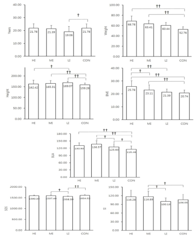
Table 2 summarizes the descriptive characteristics and QUS parameters of the athletes according to their major sporting activities. Fencing, track and field (throwing events), and softball athletes exhibited relatively high mean values of the QUS parameters. The female athletes were divided into 3 impact groups according to the body weight bearing and mechanical loading, as described in the methods section (Figure 1). Statistical estimation revealed that there was a significant difference in all the QUS parameters between the 4 groups in one-way ANOVA (P < 0.05). BUA of the HI, MI, and LI groups were significantly higher than that of the control (P < 0.01 for HI, P < 0.01 for MI, P < 0.05 for LI). SOS of the LI group was significantly lower than those of the MI (P < 0.05) and control (P < 0.01). SI of the MI group was significantly higher than those of the LI (P < 0.05) and control (P < 0.05).
Table 2
Physical characteristics and ultrasound parameters for female athletes depending on their majoring sports
| High impact group (N = 18) |
Medium impact group (N = 36) |
Low impact group (N = 16) |
|||||||
|---|---|---|---|---|---|---|---|---|---|
| Fencing (N = 3) |
Track & Field (throwing event) (N = 4) |
Weightlifting (N =11) |
Badminton (N = 8) |
Judo (N = 15) |
Softball(N = 13) | Bowling (N = 5) |
Cycling (N = 2) |
Swimming (N = 9) |
|
| Age (years) |
23.67 ± 1.15 | 23.50 ± 3.00 | 20.00 ± 3.35 | 20.75 ± 2.37 | 21.33 ± 2.85 | 21.46 ± 2.44 | 23.40 ± 4.34 | 19.50 ± 0.71 | 16.56 ± 1.94 |
| Weight (kg) |
63.8 0± 2.46 | 85.87 ± 11.16 | 63.92 ± 18.63 | 55.80 ± 3.41 | 70.92 ± 17.13 | 59.42 ± 4.46 | 62.38 ± 10.70 | 62.40 ± 0.85 | 58.92 ± 4.28 |
| Height (cm) |
167.53 ± 2.00 | 173.70 ± 6.35 | 156.93 ± 7.22 | 167.44 ± 4.26 | 165.56 ± 7.76 | 163.70 ± 6.06 | 163.98 ± 3.61 | 172.65 ± 4.88 | 171.10 ± 4.93 |
| BMI (kg/㎡) |
22.77 ± 1.33 | 28.40 ± 2.58 | 25.65 ± 5.12 | 19.90 ± 0.96 | 25.60 ± 4.33 | 22.22 ± 2.06 | 23.14 ± 3.55 | 20.95 ± 1.48 | 20.51 ± 0.87 |
| BUA (dB/MHz) |
133.38 ± 6.90 | 134.38 ± 10.10 | 129.02 ± 14.09 | 137.70 ± 11.29 | 135.76 ± 10.96 | 136.80 ± 20.40 | 128.77 ± 9.17 | 116.05 ± 14.52 | 125.96 ± 14.16 |
| SOS (m/sec) |
1560.64 ± 11.24 | 1616.07 ± 31.03 | 1589.09 ± 33.22 | 1589.39 ± 20.03 | 1594.87 ± 51.83 | 1605.27 ± 38.37 | 1571.24 ± 9.00 | 1535.34 ± 8.33 | 1556.73 ± 21.71 |
| SI | 105.67 ± 5.13 | 139.25 ± 35.78 | 110.82 ± 14.52 | 116.63 ± 12.01 | 113.93 ± 19.28 | 120.46 ± 20.98 | 105.80 ± 6.87 | 87.50 ± 12.02 | 99.78 ± 13.83 |
Figure 1.
HI, high impact group; MI, medium impact group; LI, Low impact group; CON, Control group;BMI, body mass index; BUA, broadband ultrasound attenuation; SOS, speed of sound; SI, stiffness index
Linear regression analysis for QUS parameters
Table 3 shows the results of the multiple linear regression analyses of the 3 QUS parameters. We found that participation in MI sports was significantly and positively associated with BUA compared to LI (P < 0.05) and that participation in LI sports was significantly and negatively associated with SOS compared to MI (P < 0.05). Additionally, MI was significantly and positively associated with increased SI than that of LI (P < 0.05).
Table 3
Multiple linear regression analysis for ultrasound parameters
| (A) Regression analysis for broadband ultrasound attenuation | |||||
|---|---|---|---|---|---|
| β | Standard error | Standardized β | T statistics | P value | |
| (Intercept) | -72.032 | 148.895 | -.484 | .630 | |
| Age | .373 | .516 | .086 | .723 | .472 |
| Weight | -1.174 | 1.077 | -1.119 | -1.090 | .280 |
| Height | 1.051 | .876 | .557 | 1.200 | .235 |
| Body mass index | 4.384 | 3.116 | 1.233 | 1.407 | .164 |
| High impact | -8.058 | 4.094 | -.250 | -1.968 | .053 |
| Low impact | -10.034 | 4.395 | -.299 | -2.283 | .026* |
| (B) Regression analysis for speed of sound | |||||
|---|---|---|---|---|---|
| β | Standard error | Standardized β | T statistics | P value | |
| (Intercept) | 2255.360 | 398.784 | 5.656 | .000 | |
| Age | 1.328 | 1.382 | .113 | .961 | .340 |
| Weight | 5.332 | 2.884 | 1.872 | 1.849 | .069 |
| Height | -4.113 | 2.346 | -.803 | -1.753 | .084 |
| Body mass index | -14.902 | 8.345 | -1.545 | -1.786 | .079 |
| High impact | -7.924 | 10.965 | -.091 | -.723 | .473 |
| Low impact | -30.290 | 11.772 | -.333 | -2.573 | .012* |
| (C) Regression analysis for SI | |||||
|---|---|---|---|---|---|
| β | Standard error | Standardized β | T statistics | P value | |
| (Intercept) | 112.568 | 206.083 | .595 | .554 | |
| Age | 1.307 | .714 | .219 | 1.829 | .072 |
| Weight | .592 | 1.491 | .410 | .397 | .692 |
| Height | -.256 | 1.212 | -.098 | -.211 | .834 |
| Body mass index | -1.243 | 4.313 | -.254 | -.288 | .774 |
| High impact | -1.388 | 5.666 | -.031 | -.245 | .807 |
| Low impact | -13.328 | 6.083 | -.289 | -2.191 | .032* |
Discussion
To our knowledge, this is the first cross-sectional study to compare calcaneal QUS parameters between elite female athletes and control subjects in Korean and Japanese. We aimed to analyze the calcaneal QUS results according to the impact levels of varying sporting activities (HI, MI, LI). Multiple linear regression analysis showed that increased BUA was associated with participation in MI sports and decreased SOS was associated with participation ♦in LI sports. Additionally, participation in MI sports showed a significant positive association with SI. Moreover, these associations were independent of age, height, weight, and BMI.
Our main focus was to evaluate QUS parameters of the varying sport impact level groups (HI vs. MI vs. LI). First, we would like to discuss all three parameters separately. One of the QUS parameters, BUA, reflects the attenuation of the ultrasound signal as it travels through the bone (Malavolta, et al. 2004). Multiple linear regression analysis showed that LI sports was negatively associated with BUA in comparison to MI (Table 3A). Previous reports have shown that non-weight bearing exercises, such as swimming and cycling, are associated with lower BMD, even in competitive male athletes (Barry and Kohrt 2008; Taaffe, et al. 2001; Yung, et al. 2005). Although SOS of the LI group was smallest among the groups examined in this study, including controls, previous QUS studies in male athletes indicate comparable BUA values among non-weight bearing athletes and athletes participating in weight bearing sports (Taaffe, et al. 2001; Yung, et al. 2005). Additionally, in previous BMD studies, female athletes participating in non-body weight bearing sports exhibit lower BMD than those undertaking other high impact sports (Ferry, et al. 2011; Mudd, et al. 2007). Although further studies are still necessary to validate these findings, elite female athletes participating in non-weight bearing sports or sports with low mechanical loading appear to have some problems in bone health.
SOS is another QUS parameter that refers to the speed of the ultrasound signal in bone. In the present study, we found that LI sports were associated with decreases in SOS. This finding corroborates those of previous studies showing significantly greater BMD in athletes participating in high impact sports (Bellew and Gehrig 2006; Conroy, et al. 1993; Creighton, et al. 2001). Comparable results have also been obtained from QUS studies in male athletes (Yung, et al. 2005). Because LI group included athletes undertaking non-weight bearing sports, female athletes participating in sports with non-weight bearing demonstrate a lower SOS value. However, since HI group was not associated with higher SOS value, further studies are necessary to clarify SOS value according to impact levels of varying sporting activities.
SI is a linear combination of BUA and SOS and is regarded as a robust indicator for BMD (Greenspan, et al. 1997). In the present study, we found that increases in SI were associated with MI sports. Since the coefficient for BUA is larger than that for SOS in the SI calculation formula, it is reasonable that MI was selected as a significant parameter in regression models for SOS and SI. Moreover, since weight-bearing sports increase BMD and QUS parameters in male athletes; it is not surprising to find that female athletes involved in body weight-bearing sports show higher SI.
Given that high mechanical impact is effective for bone formation, it was somewhat unexpected that HI no significantly associated with SI. It is also questionable that QUS parameters of weight-lifting athletes were not the highest among the athletes examined in this study. The BMD of male elite junior Olympic weightlifters has been reported to be significantly greater in comparison to sedentary controls (Conroy, et al. 1993). Female young adult body builders also exhibit significantly higher bone mineral content than other athletes (Heinrich, et al. 1990). On the other hand, the BMD of female adolescent weight lifters do not differ significantly from those of soccer players (Bellew and Gehrig 2006). Although participation in HI sports was not significantly associated with SI in the present study, further investigations to evaluate the effects of impact on bone quality are still needed.
Osteocytes and osteoprogenitors play a role in mature bone remodeling (Chen, et al. 2010). Mechanical stress is applied to the bone during body movements and recent studies indicate that interstitial fluid flow is a potent regulator of bone cell metabolism(Fritton and Weinbaum 2009). Stress-generated potentials across the bone tissue through fluid flow in a connected system of micropores in the bone through mechanisms such as lacunar-canalicular porosity (Piekarski and Munro 1977). Sufficient shear stress to the bone cells has been estimated with a theoretical model consisting of osteocyte process with bone matrix (Anderson and Knothe Tate 2008; Wang, et al. 2007). The results, concomitant with those of a tissue culture model (Xie, et al. 2006), suggest that high frequency (>30 Hz), low magnitude (<1 MPa) loads are sufficient for a cellular response. The level of strain on the bone has been estimated to be in the range of 0.04-0.3% (Fritton, et al. 2000), which is reportedly sufficient for bone remodeling (You, et al. 2001). These lines of evidence suggest that strain occurring in sporting activities is enough to induce bone remodeling. Furthermore, induced shear stress that exceeds optimal impact might be harmful to cells. We speculate that medium to high impact levels of sporting activities would be sufficient for bone cell-remodeling reactions to improve bone health.
Although age, weight, and BMI are frequently identified as influential variables in assessments of BMD, previous studies indicate that this is not always the case in female athletes (Arasheben, et al. 2011; Mudd, et al. 2007). In our study the associations between sport impact levels and QUS parameters in female athletes were independent of age, body weight, height and BMI. Likewise, Laabes et al. (Laabes, et al. 2008) previously reported non-significant influences of age, weight, and BMI on QUS parameters in African-American male athletes. Although a greater body mass is expected to result in higher axial loading and thus provides greater osteogenic stimulation, sporting activities such as weight-bearing, may be more influential in female athletes. Meanwhile, increased growth hormone through exercise can help bone health (Milliken, et al. 2003). However, amenorrhea, which is often experienced by female elite athletes, has been shown to reduce estrogen secretion and negatively affect bone health (Ackerman, et al. 2011). In our study, no subjects experienced amenorrhea. It was mentioned in the method.
Nevertheless, the present study also has a number of limitations that remain to be addressed. First, the criteria for determining the sport impact level groups were arbitrary defined. Although we referenced earlier studies (Groothausen, et al. 1997; Torstveit and Sundgot-Borgen 2005), and defined the categorization scheme used in this study, further revision of the classification system is still needed. We also hypothesize that including other significant parameters such as sport-specific training periods, duration, and loading in the regression models would strengthen the results. Also, bone growth and structure is markedly effected by age, age of LI group significantly lower than that of the control group. Finally, we could not draw firm conclusions regarding the causal relationships between sport impact levels and bone quality owing to the cross-sectional nature of this study. As such, further longitudinal studies examining the relationships between sporting activities and QUS parameters in young female athletes are still warranted.
In conclusion, we found that activities involving body weight impact are effective for increasing the values of QUS parameters. This finding highlights the importance of body weight bearing exercises at a young age for improving bone structure and quality to prevent osteoporosis in later life.
References
Bauer, D. C., Gluer, C. C., Cauley, J. A., Vogt, T. M., Ensrud, K. E., Genant, H. K., & Black, D. M. (1997). Broadband ultrasound attenuation predicts fractures strongly and independently of densitometry in older women. A prospective study. Study of Osteoporotic Fractures Research Group. Arch Intern Med, 157(6), 629-634.
Krieg, M. A., Cornuz, J., Ruffieux, C., Sandini, L., Buche, D., Dambacher, M. A., Hartl, F., Hauselmann, H. J., Kraenzlin, M., Lippuner, K., Neff, M., Pancaldi, P., Rizzoli, R., Tanzi, F., Theiler, R., Tyndall, A., Wimpfheimer, K., & Burckhardt, P. (2003). Comparison of three bone ultrasounds for the discrimination of subjects with and without osteoporotic fractures among 7562 elderly women. J Bone Miner Res, 18(7), 1261-1266.
Milliken, L. A., Going, S. B., Houtkooper, L. B., Flint-Wagner, H. G., Figueroa, A., Metcalfe, L. L., Blew, R. M., Sharp, S. C., & Lohman. T. G. (2003). Effects of exercise training on bone remodeling, insulin-like growth factors, and bone mineral density in postmenopausal women with and without hormone replacement therapy. Calcif Tissue Int, 72(4), 478-484.
Rhee, E. J., Oh, K. W., Lee, W. Y., Kim, S. W., Oh, E. S., Baek, K. H., Kang, M. I., Park, C. Y., Choi, M. G., Yoo, H. J., & Park, S. W. (2004). Age, body mass index, current smoking history, and serum insulin-like growth factor-I levels associated with bone mineral density in middle-aged Korean men. J Bone Miner Metab, 22(4), 392-398.