Comparison of knee valgus motion in African American male and female collegiate athletes
Article information
Abstract
Knee joint valgus is often implicated as a hazardous position for the anterior cruciate ligament (ACL) and has been linked to ACL injury risk. Although several groups have previously examined racially diverse cohorts to determine if there are predispositions for injury, there is little evidence found that explore sex differences in valgus angles in African American athletes. The purpose of this study was to identify the difference of the knee valgus angles between African American male (AAM) and female (AAF) student-athletes. Eighty-five National Collegiate Athletic Association, Division II African American male (n=36) and female (n=49) student-athletes participated in this study. Subjects dropped directly down off a box and immediately performed a maximum vertical jump. Valgus and flexion angles were analyzed and calculated based on the relationship of the tangent for each image. Knee valgus angles were significantly different between AAM and AAF athletes both at initial contact and maximum valgus displacement (p<.05). Significant differences in knee flexion angle was observed with the AAM having greater Initial Contact knee flexion (p<.05). The current data indicates that sex differences, specifically increased knee valgus angles, exist during jumping and landing. Neuromuscular training programs should be designed to specifically address excessive valgus knee motion and improve landing knee flexion among these athletes.
Introduction
Anterior cruciate ligament (ACL) injury is a critical and one of the most common sports injuries that results from the changing direction at speed while playing a sport. According to the previous researches, female athletes are currently reported to be four to six times more likely to sustain a sports related non-contact ACL injury than male athletes (Malone, Hardaker, Garrett, Feagin, & Bassett, 1995; Marshall, Padua, Shultz, & McGrath, 2007). Arendt and Dick established that females are at a greater risk for ACL injury compared to males that participated in basketball and soccer (Arendt & Dick, 1995; Webster & Gribble, 2010).
Structural differences exist between sex and races which may be associated with biomechanics that relate to increased injury risk (Al-Saeed, Brown, Athyal, & Sheikh, 2013; Rizzo, Holler, & Bassett, 2001; Shelbourne, Gray, & Benner, 2007). Everhart et al. compared the intercondylar notch widths by race and sex of 90 males and 79 females. The investigators found that women had 17.1% narrower intercondylar notch width and 5.2% higher percent stenosis than men. African American males (AAM) were found to have 8.3% narrower notch width than Caucasian men with no difference between women of both races (Everhart, Flannifan, & Chaudhari, 2014).
Racial differences in risk factors may help explain variations in the natural history of knee diseases (Chang et al., 2010) and athletic injuries (Davis, Shelbourne, & Klootwyk, 1999; Gianotti, Marshall, Hume, & Bunt, 2009; Shelbourne et al., 2007). Chang et al. undertook a study to determine the frequency of varus and valgus thrust in African Americans and Caucasians (Chang et al., 2010). The authors concluded that compared with Caucasians, African Americans had lower odds of varus thrust (Odd Ratio, 0.50) and greater odds of valgus thrust (Odd Ratio, 1.69) which may explain the difference between patterns of osteoarthritis involvement in the knee (Chang et al., 2010).
Injury risk factors have previously been examined in racially diverse cohorts to determine if there are predispositions for injury (Wren et al., 2012). However, there are limited data that evaluate African American female (AAF) athletes relative to injury and injury risk. Also, these researches compared static knee alignment among different races. Health care professionals continue to face the challenge of preventing ACL injuries in female collegiate athletes and rehabilitating those who have ACL injuries (Webster & Gribble, 2010). By understanding the mechanisms of injury in sport and physical activities, it is crucial to investigate them during kinetic movement. Hewett et al. (Ford, Myer, Toms, & Hewett, 2005) conducted a prospective study that investigated biomechanical measures of neuromuscular control and valgus loading of the knee and concluded that knee motion and knee loading during a landing task are predictors of ACL injury risk in female athletes. Paterno et al (Paterno et al., 2010) reported that altered neuromuscular control of the hip and knee during a dynamic landing task and postural stability deficits after anterior cruciate ligament reconstruction (ACLR) are predictors of a second ACL injury after an athlete is released to return to sport. Although several groups have previously examined sex differences in knee joint valgus (abnormal outward angulation of the distal segment of joint) during a drop vertical jump (Ford, Myer, & Hewett, 2003; Ford et al., 2005; Hewett et al., 2005), there is little evidence found that explores sex differences in knee joint kinematics, or ACL injury rates, in various races. Even though the percentage of AAF student-athletes participating in NCAA sports has increased from 9.4 to 11.6 percent over the past years (Erin, 2010), they are under-represented in studies related to the identification of risk and prevention of ACL injuries that directly identify kinematic sex specific differences in African American athletes.
There appears to be a gap in research relating to knee valgus kinematics and sex differences among African American athletes when performing a landing maneuver. Therefore, the purpose of this study was to identify if injury risk factors, specifically knee valgus angles, differ between AAM and AAF student-athletes in a Division II Historically Black College and University.
Methods
Participants
Eighty-five National Collegiate Athletic Association (NCAA) Division II African American male and female student-athletes volunteered to participate in this study (Female: n=49, Males: n=36). All athletes were cleared by team physicians through pre-participation physical and were afforded an opportunity to volunteer for this study. Athletes not cleared for participation and those athletes with prior ACL injury/-reconstruction were excluded from this study. Any volunteers younger than age 18 were also excluded. Each participant signed an informed consent prior to participation. The protocol for this study was approved by Albany State University and Rocky Mountain University of Health Professions Institutional Review Boards.
Procedures
Two members from the Sports Medicine and Re-conditioning Team (SMART) used a wall-mounted height rod (Stadi-O-Meter, Novel Products, Inc., Rockton, Illinois) to measure height, a Tanita WB – 3000 Digital Scale (Tanita Corporation, Japan) to measure weight, and an OMRON HBR-360 Body fat Analyzer (Omron Healthcare Inc., Vernon Hills, Illinois) to measure body fat. Clinic-based tibia lengths were measured with the subject standing with knees extended in anatomical position. A standard measuring tape was used to measure the distance between the lateral knee joint line and the lateral malleolus (Myer, Ford, & Hewett, 2010).
A box (31cm high), centered on the frontal camera view and approximately 30cm off center in the sagittal plane, was used for the drop vertical jump (DVJ). The subjects were instructed to stand on top of the box with their feet positioned 35cm apart (Figure 1) (Ford, Myer, & Hewett, 2003).
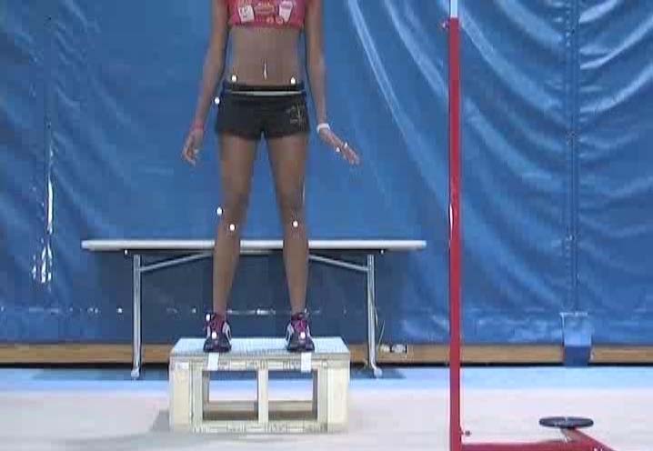
Drop vertical jump box
Two-dimensional frontal and sagittal plane data was captured using standard video cameras. Cameras were leveled at 80 cm in height and perpendicular in the sagittal and frontal planes. The box (31 cm high) used for the drop-vertical jump (DVJ) was centered on the frontal camera view and approximately 30 cm off center of sagittal plane view away from the camera in the frontal plane position.
To perform the DVJ, a standardized target was placed overhead, equal to the subject’s maximal touch height during a countermovement vertical jump. Once prepared, the subjects dropped directly down off the box and immediately performed a maximum vertical jump, raising both arms towards the overhead target. Subjects were allowed one to three practice trials to ensure they were able to demonstrate adequate understanding and ability to perform the instructed test maneuver (Myer et al., 2010). Knee kinematic data was collected in the frontal and sagittal plane using a standard video camera (Canon XL2 3CCD DV) with an effective sampling rate of 60Hz. The cameras were positioned at a height of 80cm and leveled perpendicular to each other in the frontal and sagittal planes (Myer et al., 2010). The investigator prepared each subject with eight retro-reflective markers (anterior superior iliac spine, greater trochanter, lateral knee joint line, and lateral malleolus on both extremities) using a double-sided tape and adhesive spray (Myer, Ford, Palumbo, & Hewett, 2005). These marked feet positions were used in order to replicate the methods in previous studies (Ford, Myer, & Hewett, 2003) and to allow medial knee motion to occur during landing. For each participant, three trials were collected on one continuous video file. The video view of the third jump was used for each participant and collected for synchronization into the motion analysis software.
Two videos that synchronized with Dartfish Motion Analysis software (Alpharetta, GA) were collected and imported to freeware software ImageJ (Rasband W S, ImageJ, US National Institutes of Health, Bethesda, Maryland, USA, http://rsb.info.nih.gov/ij/, 1997-2009) to digitize the images. Four image files were captured in the following order: I1-frontal plane view with frame prior to initial contact, I2-frontal plane view of frame with knee in maximum medial (valgus) position, I3-sagittal plane view with frame prior to initial contact, I4-sagittal plane view of frame with the knee in maximum flexion position and named in a standard structure for each subject (i.e., SubjectX I1, SubjectX I2, SubjectX I3, SubjectX I4) (Myer et al., 2010). Valgus and flexion angles were calculated based on the relationship of the tangent for each image (Figure 2).
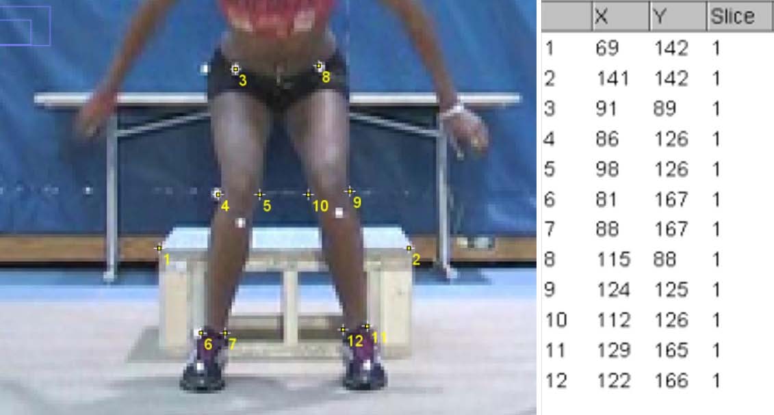
Valgus and flexion angle calculations
Using a double sided tape and adhesive spray, the investigator attached each subject with eight retro-reflective markers placed on the ASIS, greater trochanter, lateral knee joint line, and lateral malleolus of both extremities. The digitized hip, knee and ankle marker coordinates were exported to excel. Valgus and flexion angles were calculated based on the relationship of the tangent for each image.
Valgus angle, Knee flection angle (θ)
Statistical Analysis
An independent t-test was used to identify sex differences between AAF and AAM groups, using a significance level of 0.05, and power .80. In order to achieve 80% power (α level=0.05) a minimum of 23 subjects was required in each group (male, female), for 2-dimensional valgus angle calculations. This was based on means and SDs differences Schmitz et al. (2009) identified in knee valgus alignment among post pubertal males 9.2 (9.4) and females 15.5 (8.7) (Schmitz, Schultz,&Nguyen, 2009).
Results
Participant characteristics are shown in Table 1. All kinematic variables for both AAM and AAF while performing the drop vertical jump are shown in Table 2 and Table 3.

Descriptive data on participants (African American Male and Female)

Valgus angles at each stage of the landing (Frontal view)

ROM angles during the drop vertical jump (Lateral view)
Knee valgus angles were significantly different between African American male and female athletes both at initial contact and maximum valgus displacement. Specifically, AAFs displayed greater knee valgus angle at initial contact (IC), (p<0.001) and had greater maximum knee valgus angle (p<0.001). Significant differences in knee flexion angle was observed between AAF and AAM groups with the AAM having greater IC knee flexion at (p=0.02). However, there were no significant differences in left or right valgus ROM between AAM and AAF groups.
Discussion
The purpose of this study was to investigate the risk factors associated with knee injuries in African American collegiate athletes. The drop vertical jumps were performed and analyzed to investigate the kinetic and mechanical differences during active movement. We found elevated risk factors in valgus angles among the AAFs that were statistically different when compared to AAM athletes. We observed differences at both initial contact and at maximum valgus displacement. We also observed significant differences in knee flexion angles at initial contact.
Generally, injury patterns in athletes have been suggested to be more sport-specific than sex-specific (Ristolainen, Heinonen, Waller, Kujala, & Kettunen, 2009). However sport injury studies continue to highlight and investigate sex differences that focus particularly on knee injuries (Arendt & Dick, 1995; Ristolainen et al., 2009). Racial disparities and sex differences have been demonstrated among specific injuries including fractures, Achilles tendon ruptures, knee disabilities, sprains, and strains (Everhart et al., 2014; Hansen, Aagaard, & Magnusson, 2012; Raikin, Garras, & Krapchev, 2013; Wren et al., 2012). Everhart et al. concluded that women have narrower femoral notches than men and African American men have narrower notches than Caucasian men and no difference was discovered between AAF and Caucasian females. Ethnically, Wren et al. noted boys and girls of European descent had double the fracture risk of children from other backgrounds suggesting a predisposition to fractures (Wren et al., 2012). Peak failure stress in Iliopsoas tendon was higher in AA than Caucasians.
We believe that the risk factors during the sports events might impose the meaningful outcomes and should be investigated during the dynamic move. Dynamic movement along with structural, racial, and sexual differences, might impact whether gender differences existed in knee valgus kinematics in AA collegiate athletes when performing a landing maneuver. To our knowledge, the current report provides the first assessment of biomechanical or neuromuscular imbalances in focused on African American females versus males during the drop vertical jump. This study found a significant effect of sex on frontal plane landing mechanics which could account for differences in risk for athletic injury.
Knee joint valgus is often implicated as a hazardous position for the ACL and has been linked to ACL injury risk (Ford et al., 2003; Ford et al., 2005; McLean, Huang, Su, & van den Bogart, 2004). Our results revealed AAF athletes land in greater knee valgus than AAM athletes. Three basic theories (anatomical, hormonal, and biomechanical) have been established as the underlying potential mechanism for the injury rate differences among male and female athletes (Ford et al., 2003). Numerous studies support the notion that females perform high demand athletic movements differently than males and in a way that predisposes them to higher knee joint stress (McLean et al., 2007; Weinhandl, Joshi, & O’Conner, 2010). Yasar et al. (Yasar, Behzat, Cengiz, Akin, & Feza, 2004) compared the landing maneuvers between male and female college volleyball players. Females demonstrated significantly lower knee and hip flexion angles compared to their male counterparts and authors concluded that female volleyball players initiate different lower extremity mechanics during landings than that of males (Yasar et al., 2004). Previous sports related studies have compared injury risk and injury types between male and female athletes among different sports (Ristolainen, Heinonen, Waller, Kujala, & Kettunen, 2009) however, there were no studies found reporting landing maneuver differences among AAM and AAF athletes.
In this study a significant difference in knee flexion at initial contact was observed between AAF and AAM groups with the AAM having greater knee flexion at initial contact. A valgus collapse of the knee joint is commonly seen with non-contact ACL injuries and often occurs with knee near full extension (between 0° and 30°), and external rotation with the foot planted during a deceleration movement (Myer, 2011). Video studies by Krosshaug et al (Krosshaug, Nakamae, & Boden, 2007) revealed dynamic valgus as the most common ACL injury mechanism for female basketball and handball players. In addition Krosshaug et al. (Krosshaug et al., 2007) found relative risk valgus collapse during ACL injury increased 5.3 times in female basketball athletes when compared with male basketball athletes. Similar results by Ford et al. (Ford et al., 2003) determined sex-related differences in knee valgus motion in high school athletes. Using two different techniques knee valgus motion was obvious in female athletes compared to male athletes, one method involved calculating the distance between the right and left knee after dropping from a box before performing a maximum vertical jump (Ford et al., 2005). Females often demonstrate more knee valgus motion at the point of maximum valgus. Russell et al. (Russell, Palmieri, Zinder, & Ingersoll, 2006) revealed women were in knee valgus immediately upon contact with the ground (e.g., before forces were transferred from the ground to the body), while men landed in knee varus (inward angulation of the distal segment of a joint). Landing strategies used by athletes are important and these findings implied that women may be conditioned with an ineffective and potentially hazardous landing strategy (Russell et al., 2006).
According to Ford et al. observed increases in knee motion (valgus knee motion and valgus angle) implies altered muscular control of the lower extremity in the frontal plane (Ford et al., 2003). The authors also advocate limiting the valgus position of the knee during landing could reduce strain on the ACL and in turn reduce the number of noncontact ACL injuries (Ford et al., 2003). According to the previous data, it is considered that dynamic neuromuscular training may be beneficial in African American female athletes to decrease the risk of ACL injuries (Hewett et al., 2005; Myer et al., 2005). The entire population of female athletes could potentially benefit from injury prevention training programs prior to participation in sports. However, those athletes that demonstrate poor dynamic knee stability may gain an increased benefit from training. Myer et al. (Myer et al., 2005) developed a new method to identify athletes at high risk for ACL injury using clinic-based measurements and freeware computer analysis that was employed in the current investigation. Future investigation may benefit from the inclusion of these clinic-based measurements to identify high-risk athletes and develop neuromuscular training programs that may decrease their risk to levels parallel to males. The current research findings indicate a mechanism (increased dynamic knee valgus and reduced knee flexion) that AAF athletes demonstrated which may place them at higher risk than their male counterparts for ACL injury. Due to the increase knee valgus demonstrated during jump maneuvers, prevention measures, such as a neuromuscular training program, might be of particular benefit for this population. In addition, comparison studies among races might enlarge the impact of this research.
Conclusion
The current data indicates that sex differences between AAF and AAM, specifically increased knee valgus angles exist during jumping and landing. This difference may indicate an increased risk for noncontact ACL injuries among AAF athletes when compared to AAM athletes. Neuromuscular training programs should be designed to specifically address excessive valgus knee motion and improve landing knee flexion among these athletes. Correction of neuromuscular imbalances is important for both the optimal biomechanics of athletic movements and reduction of knee injury incidence. Further study on the effects of neuromuscular training on knee valgus among AAF athletes is important for the advancement of injury prevention and safe participations in athletics and is particularly important at Division II Historically Black Colleges and Universities this is the first reference to HBCUs noted.
References
Al-Saeed, O., Brown, M., Athyal, R., & Sheikh, M. (2013). Association of femoral intercondylar notch morphology, width index and the risk of anterior cruciate ligament injury. Knee Surg Sports Traumatol Arthrosc, 21(3), 678-682. doi:10.1007/s00167-012-2038-.
Al-Saeed O., Brown M., Athyal R., et al, Sheikh M.. 2013;Association of femoral intercondylar notch morphology, width index and the risk of anterior cruciate ligament injury. Knee Surg Sports Traumatol Arthrosc 21(3):678–682. 10.1007/s00167-012-2038-y.Arendt, E., & Dick, R. (1995). Knee injury patterns among men and women in collegiate basketball and soccer: NCAA data and review of literature. American Journal of Sports Medicine, 23(6), 694-701..
Arendt E., et al, Dick R.. 1995;Knee injury patterns among men and women in collegiate basketball and soccer: NCAA data and review of literature. American Journal of Sports Medicine 23(6):694–701.Chang, A., Hochberg, M., Dunlop, D., Chimiel, J., Nevitt, M., Hayes, K., . . . Sharma, L. (2010). Frequency of varus and valgus thrust and factors associated with thrust presence in persons with or at higher risk of developing knee osteoarthritis. Arthritis Rheum, 62(5), 1403-1411..
Chang A., Hochberg M., Dunlop D., Chimiel J., Nevitt M., Hayes K., . . . Sharma L.. 2010;Frequency of varus and valgus thrust and factors associated with thrust presence in persons with or at higher risk of developing knee osteoarthritis. Arthritis Rheum 62(5):1403–1411. 10.1002/art.27377.Davis, T. J., Shelbourne, K. D., & Klootwyk, T. E. (1999). Correlation of the intercondylar notch width of the femur to the width of the anterior and posterior cruciate ligaments. Knee Surg Sports Traumatol Arthrosc, 7(4), 209-214. doi:10.1007/s00167005015.
Davis T. J., Shelbourne K. D., et al, Klootwyk T. E.. 1999;Correlation of the intercondylar notch width of the femur to the width of the anterior and posterior cruciate ligaments. Knee Surg Sports Traumatol Arthrosc 7(4):209–214. 10.1007/s001670050150.Everhart, J., Flannifan, D., & Chaudhari, A. (2014). Anteromedical ridging of the femoral intercondylar notch: An anatomic study of 170 archival skeletal specimens. Knee Surg Traumatol Arthrosc, 22(1), 80-87..
Everhart J., Flannifan D., et al, Chaudhari A.. 2014;Anteromedical ridging of the femoral intercondylar notch: An anatomic study of 170 archival skeletal specimens. Knee Surg Traumatol Arthrosc 22(1):80–87. 10.1007/s00167-012-2282-1.Ford, K. R., Myer, G. D., & Hewett, T. E. (2003). Valgus knee motion during landing in high school female and male basketball players. Med Sci Sports Exerc, 35(10), 1745-1750. doi: 10.1249/01.MSS.0000089346.85744.D.
Ford K. R., Myer G. D., et al, Hewett T. E.. 2003;Valgus knee motion during landing in high school female and male basketball players. Med Sci Sports Exerc 35(10):1745–1750. 10.1249/01.mss.0000089346.85744.d9.Ford, K. R., Myer, G. D., Toms, H. E., & Hewett, T. E. (2005). Gender differences in kinematics of unanticipated cutting in young athletes. Medicine and Science in Sports and Exercise, 37, 124-129.
. Ford K. R., Myer G. D., Toms H. E., et al, Hewett T. E.. 2005;Gender differences in kinematics of unanticipated cutting in young athletes. Medicine and Science in Sports and Exercise 37:124–129.Gianotti, S. M., Marshall, S. W., Hume, P. A., & Bunt, L. (2009). Incidence of anterior cruciate ligament injury and other knee ligament injuries: A national population-based study. J Sci Med Sport, 12(6), 622-627. doi:10.1016/j.jsams.2008.07.00.
Gianotti S. M., Marshall S. W., Hume P. A., et al, Bunt L.. 2009;Incidence of anterior cruciate ligament injury and other knee ligament injuries: A national population-based study. J Sci Med Sport 12(6):622–627. 10.1016/j.jsams.2008.07.005.Hansen, P., Aagaard, P., & Magnusson, S. (2012). Biomechanical properties of isolated fascicles of the iliopsoas and Achilles tendons in African American and Caucasian men. Ann Anat., 194(5), 457-460..
Hansen P., Aagaard P., et al, Magnusson S.. 2012;Biomechanical properties of isolated fascicles of the iliopsoas and Achilles tendons in African American and Caucasian men. Ann Anat. 194(5):457–460.Hewett, T. E., Myer, G. D., Ford, K. R., Heidt, R., Jr., Colosimo, A. J., & McLean, S. G. (2005). Biomechanical measures of neuromuscular control and valgus loading of the knee predict anterior cruciate ligament injury risk in female athletes: A prospective study. American Journal of Sports Medicine, 33(4), 492-501.
Hewett T. E., Myer G. D., Ford K. R., Heidt R. Jr., Colosimo A. J., et al, McLean S. G.. 2005;Biomechanical measures of neuromuscular control and valgus loading of the knee predict anterior cruciate ligament injury risk in female athletes: A prospective study. American Journal of Sports Medicine 33(4):492–501. 10.1177/0363546504269591.Krosshaug, T., Nakamae, A., & Boden, B. P. (2007). Mechanisms of anterior cruciate ligament injury in basketball: Video analysis of 39 cases. American Journal of Sports Medicine, 35, 359-367.
Krosshaug T., Nakamae A., et al, Boden B. P.. 2007;Mechanisms of anterior cruciate ligament injury in basketball: Video analysis of 39 cases. American Journal of Sports Medicine 35:359–367.Malone, T., Hardaker, W., Garrett, W., Feagin, J., & Bassett, F. (1995). Relationship of gender to anterior cruciate ligament injuries in intercollegiate basketball players. Journal of Southern Orthopaedic Association, 2(1), 36-39..
Malone T., Hardaker W., Garrett W., Feagin J., et al, Bassett F.. 1995;Relationship of gender to anterior cruciate ligament injuries in intercollegiate basketball players. Journal of Southern Orthopaedic Association 2(1):36–39.Marshall, S., Padua, D., Shultz, S., & McGrath, M. (2007). The incidence of ACL injury. Understanding and Preventing Non-Contact ACL Injuries. (pp. 5-29). Champaign: Human Kinetics.
Marshall S., Padua D., Shultz S., et al, McGrath M.. 2007. The incidence of ACL injury. Understanding and Preventing Non-Contact ACL Injuries p. 5–29. Champaign: Human Kinetics.McLean, S. G., Fellin, R. E., Suedekum, N., Calabrese, G., Passerallo, A., & Joy, S. (2007). Impact of fatigue on gender-based high-risk landing strategies. Medicine and Science in Sports and Exercise, 39(3), 502-514..
McLean S. G., Fellin R. E., Suedekum N., Calabrese G., Passerallo A., et al, Joy S.. 2007;Impact of fatigue on gender-based high-risk landing strategies. Medicine and Science in Sports and Exercise 39(3):502–514. 10.1249/mss.0b013e3180d47f0.McLean, S. G., Huang, X., Su, A., & van den Bogart, A. J. (2004). Sagittal plane biomechanics cannot injure the ACL during sidestep cutting. Clinical Biomechanics, 19, 828-838.
. McLean S. G., Huang X., Su A., et al, van den Bogart A. J.. 2004;Sagittal plane biomechanics cannot injure the ACL during sidestep cutting. Clinical Biomechanics 19:828–838. 10.1016/j.clinbiomech.2004.06.006.Myer, G. (2011). Three-Dimensional Motion Analysis Validation of a Clinic-Based Nomogram Designed to Identify High ACL Injury Risk in Female Athletes. The Physician and Sportsmedicine, 39(1), 19-28..
Myer G.. 2011;Three-Dimensional Motion Analysis Validation of a Clinic-Based Nomogram Designed to Identify High ACL Injury Risk in Female Athletes. The Physician and Sportsmedicine 39(1):19–28. 10.3810/psm.2011.02.1838.Myer, G., Ford, K., Palumbo, J., & Hewett, T. (2005). Neuromuscular Training Improves Performance and Lower-Extremity Biomechanics in Female Athletes. Journal of Strength and Conditioning Research, 19(1), 51-60..
Myer G., Ford K., Palumbo J., et al, Hewett T.. 2005;Neuromuscular Training Improves Performance and Lower-Extremity Biomechanics in Female Athletes. Journal of Strength and Conditioning Research 19(1):51–60. 10.3810/psm.2011.02.1858.Myer, G. D., Ford, K. R., & Hewett, T. (2010). New method to identify athletes at high risk of ACL injury using clinic-based measurements and freeware computer analysis. Br J Sports Med., doi: 10.1136/bjsm.2010.072843
. Myer G. D., Ford K. R., et al, Hewett T.. 2010;New method to identify athletes at high risk of ACL injury using clinic-based measurements and freeware computer analysis. Br J Sports Med 10.1136/bjsm.2010.072843.Paterno, M. V., Schmitz, L., Ford, K., Rauh, M., Myer, G., Huang, B., & Hewett, T. (2010). Biomechanical Measures During Landing and Postural Stability Predict Second Anterior Cruciate Ligament Injury After Anterior Cruciate Ligament Reconstruction and Return to Sport. Am J Sports Med, 38(10), 1968-1978.
Paterno M. V., Schmitz L., Ford K., Rauh M., Myer G., Huang B., et al, Hewett T.. 2010;Biomechanical Measures During Landing and Postural Stability Predict Second Anterior Cruciate Ligament Injury After Anterior Cruciate Ligament Reconstruction and Return to Sport. Am J Sports Med 38(10):1968–1978. 10.1177/0363546510376053.Raikin, S., Garras, D., & Krapchev, P. (2013). Achilles tendon injuries in a United States population. Foot Ankle Int, 34(4), 475-480..
Raikin S., Garras D., et al, Krapchev P.. 2013;Achilles tendon injuries in a United States population. Foot Ankle Int 34(4):475–480. 10.1177/1071100713477621.Ristolainen, L., Heinonen, A., Waller, B., Kujala, U. M., & Kettunen, J. A. (2009). Gender differences in sport injury risk and types of injuries: A retrospective twelve-month study on cross-country skiers, swimmers, long-distance runners and soccer players. Journal of Sports Science and Medicine, 8, 443-451.
. Ristolainen L., Heinonen A., Waller B., Kujala U. M., et al, Kettunen J. A.. 2009;Gender differences in sport injury risk and types of injuries: A retrospective twelve-month study on cross-country skiers, swimmers, long-distance runners and soccer players. Journal of Sports Science and Medicine 8:443–451.Rizzo, M., Holler, S. B., & Bassett, F. H., 3rd. (2001). Comparison of males’ and females ratios of anterior-cruciate-ligament width to femoral-intercondylar-notch width: A cadaveric study. Am J Orthop (Belle Mead NJ), 30(8), 660-664..
Rizzo M., Holler S. B., et al, Bassett, F. H. 3rd.. 2001;Comparison of males’ and females ratios of anterior-cruciate-ligament width to femoral-intercondylar-notch width: A cadaveric study. Am J Orthop (Belle Mead NJ) 30(8):660–664.Russell, K. A., Palmieri, R. M., Zinder, S. M., & Ingersoll, C. D. (2006). Sex Differences in Valgus Knee Angle During a Single-Leg Drop Jump. Journal of Athletic Training, 41(2), 166-171..
Russell K. A., Palmieri R. M., Zinder S. M., et al, Ingersoll C. D.. 2006;Sex Differences in Valgus Knee Angle During a Single-Leg Drop Jump. Journal of Athletic Training 41(2):166–171.Schmitz, R., Schultz, S., & Nguyen, A. D. (2009). Dynamic Valgus Alignment and functional Strength in Males and Females During Maturation. Journal of Athletic Training, 44(1), 26-32..
Schmitz R., Schultz S., et al, Nguyen A. D.. 2009;Dynamic Valgus Alignment and functional Strength in Males and Females During Maturation. Journal of Athletic Training 44(1):26–32. 10.4085/1062-6050-44.1.26.Shelbourne, K. D., Gray, T., & Benner, R. W. (2007). Intercondylar notch width measurement differences between African American and white men and women with intact anterior cruciate ligament knees. Am J Sports Med, 35(8), 1304-1307. doi: 10.1177/036354650730006.
Shelbourne K. D., Gray T., et al, Benner R. W.. 2007;Intercondylar notch width measurement differences between African American and white men and women with intact anterior cruciate ligament knees. Am J Sports Med 35(8):1304–1307. 10.1177/0363546507300060.Webster, K. A., & Gribble, P. A. (2010). Time to Stabilization of Anterior Cruciate Ligament Reconstructed Versus Healthy Knees in National Collegiate Athletic Association Division I Female Athletes. Journal of Athletic Training, 45(6), 580-585..
Webster K. A., et al, Gribble P. A.. 2010;Time to Stabilization of Anterior Cruciate Ligament Reconstructed Versus Healthy Knees in National Collegiate Athletic Association Division I Female Athletes. Journal of Athletic Training 45(6):580–585. 10.4085/1062-6050-45.6.580.Weinhandl, J. T., Joshi, M., & O’Conner, K. (2010). Gender Comparisons Between Unilateral and Bilateral Landings. Journal of Applied Biomechanics, 26, 444-453.
. Weinhandl J. T., Joshi M., et al, O’Conner K.. 2010;Gender Comparisons Between Unilateral and Bilateral Landings. Journal of Applied Biomechanics 26:444–453. 10.1123/jab.26.4.444.Wren, T. A., Shepherd, J. A., Kalkwarf, H. J., Zemel, B. S., Lappe, J. M., Oberfield, S., . . . Gilsanz, V. (2012). Racial disparity in fracture risk between white and nonwhite children in the United States. J Pediatr, 161(6), 1035-1040. doi: 10.1016/j.jpeds.2012.07.05.
Wren T. A., Shepherd J. A., Kalkwarf H. J., Zemel B. S., Lappe J. M., Oberfield S., . . . Gilsanz V.. 2012;Racial disparity in fracture risk between white and nonwhite children in the United States. J Pediatr 161(6):1035–1040. 10.1016/j.jpeds.2012.07.054.Yasar, S., Behzat, B. K., Cengiz, H., Akin, S., & Feza, K. (2004). Comparison of landing maneuvers between male and female college volleyball players. Clinical Biomechanics, 19(6), 622-628.
Yasar S., Behzat B. K., Cengiz H., Akin S., et al, Feza K.. 2004;Comparison of landing maneuvers between male and female college volleyball players. Clinical Biomechanics 19(6):622–628.Zgon, E. (2010). NCAA student athlete ethnicity report. Indianapolis, IN: National Collegiate Athletic Association.
Zgon E.. 2010. NCAA student athlete ethnicity report Indianapolis, IN: National Collegiate Athletic Association;